


Quick Links
For Patients
© Copyright 2026 American TelePhysicians. All rights reserved.
Several conditions can affect the heart valves like;
Several conditions can affect the heart valves like;
Types of valvular disorders are based on the type of valve involved, which can be;
Types of valvular disorders are based on the type of valve involved, which can be;
Valvular heart disease affects about 2.5% of the population In the United States. The frequency of these disorders increases as the person ages, and it is prevalent in 13 % of 75 year-olds in the United States. The most common cause of valve diseases in developing countries is rheumatic disease, which makes up to 65% of valve disorders. Aortic stenosis and regurgitation are affected mainly by age, and their prevalence is 12.4% and 13% in the age group of 55-80 years, respectively. Mitral stenosis is almost always caused by rheumatic heart disease, and it is prevalent in 0.1% of people in the US. 9% of people over 75 years have mitral regurgitation due to aging.
Valvular heart disease affects about 2.5% of the population In the United States. The frequency of these disorders increases as the person ages, and it is prevalent in 13 % of 75 year-olds in the United States. The most common cause of valve diseases in developing countries is rheumatic disease, which makes up to 65% of valve disorders. Aortic stenosis and regurgitation are affected mainly by age, and their prevalence is 12.4% and 13% in the age group of 55-80 years, respectively. Mitral stenosis is almost always caused by rheumatic heart disease, and it is prevalent in 0.1% of people in the US. 9% of people over 75 years have mitral regurgitation due to aging.
Having one or more of the following risk factors puts you at an increased chance of having a valvular disease:
Having one or more of the following risk factors puts you at an increased chance of having a valvular disease:
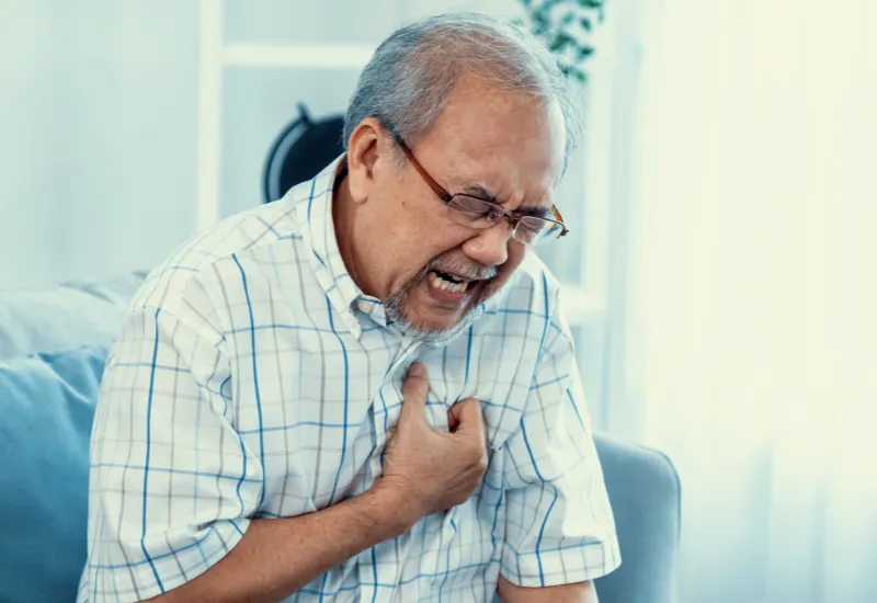
The following symptoms may point towards a heart valve disease, and you should plan to consult a physician.
The following symptoms may point towards a heart valve disease, and you should plan to consult a physician.
Your doctor will inquire about the symptoms and the previous medical records to diagnose the disorder. He will examine with particular emphasis on the heart examination using a stethoscope for hearing the heart sounds and murmurs to determine valvular heart disease. The following tests can be used:
Your doctor will inquire about the symptoms and the previous medical records to diagnose the disorder. He will examine with particular emphasis on the heart examination using a stethoscope for hearing the heart sounds and murmurs to determine valvular heart disease. The following tests can be used:
Some other disorders may present like valvular heart disease that needs to be excluded from making an efficient diagnosis:
Some other disorders may present like valvular heart disease that needs to be excluded from making an efficient diagnosis:
Children with valvular defects due to congenital heart disease can be cured entirely in infancy by surgical repair. Adults with valvular heart disease are treated differently depending on the severity of their condition. You could need to be watched or need medicine or surgery.
Minor heart defects may only necessitate periodic health checks with your specialist to help that your illness does not deteriorate. Inquire with your physician about how frequently you should be seen.
Medications
Medications that assist the heart to perform better can be used to cure some moderate heart abnormalities. Several classes of medicines have been used depending on the type of the condition and the symptoms needed to be controlled, like anticoagulants (Heparin, Warfarin) are used to avoid blood clots or Beta Blockers (Atenolol, Propranolol), and Calcium Channel Blockers (Amlodipine, Diltiazem ) are used to manage an abnormal heart rhythm. Diuretics (Furosemide, Bumetanide) are used to treat edema associated with heart disease.
Surgical repair
You may need surgical correction of the heart valves, including tightening the ring at the base of the valve, separating the fused leaflets of the valves, etc. Valve replacement procedure can be performed using catheterization or open-heart surgery.
Prognosis depends on the valve involved and how well the disease is managed. Some are managed well by medications and follow-ups, while others are cured using surgeries. For untreated patients, the outlook is poor.
Prognosis depends on the valve involved and how well the disease is managed. Some are managed well by medications and follow-ups, while others are cured using surgeries. For untreated patients, the outlook is poor.
Adopting the following lifestyle changes may prevent you from developing valvular heart disease.
Adopting the following lifestyle changes may prevent you from developing valvular heart disease.
Our clinical experts continually monitor the health and medical content posted on CURA4U, and we update our blogs and articles when new information becomes available. Last reviewed by Dr.Saad Zia on June 02, 2023.
Global epidemiology of valvular heart disease | Nature Reviews Cardiology
https://www.nature.com/articles/s41569-021-00570-z
Heart Valve Disease | American Heart Association
https://www.heart.org/en/health-topics/heart-valve-problems-and-disease


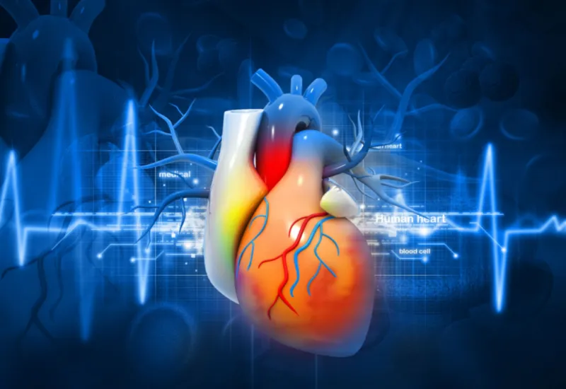
The disorder involving your heart valves is referred to as valvular heart disease. Your heart is a muscular organ located in the center of the chest. Its function is to pump blood with oxygen and nutrients throughout the body and bring back the blood containing carbon dioxide taken to the lungs to exit the body. It consists of four chambers; the upper two are atria, and the lower two are called ventricles. These chambers are separated by valves that allow the blood to flow in one direction only to avoid mixing the blood to prevent disruption of heart function. Your heart contains four valves. The two are between the atria and ventricle, while the other two are at the commencement of main blood vessels taking blood away from the heart to the body and the lungs, i.e., the aorta and pulmonary artery.
Any condition affecting these valves results in the disruption of heart function, and so the symptoms depend on the valve type involved. They are cured either with medicines, surgery, or both.