

X-Ray Wrist 2 Views-Left
X-Ray
Upper Extremities/Limbs
Wrist
2 views

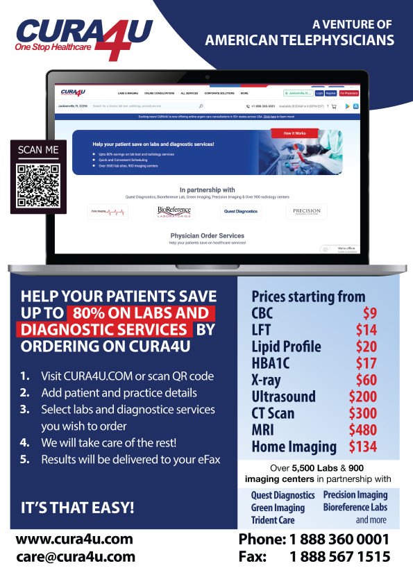
Green Imaging, a leading provider of advanced medical imaging services, in partnership with CURA4U brings affordable and high-quality diagnostic imaging solutions to individuals seeking comprehensive healthcare. With a commitment to making advanced medical imaging accessible, Green Imaging offers a diverse range of services through CURA4U, empowering patients to take control of their health and well-being.
Green Imaging boasts a network of imaging centres equipped with state-of-the-art technology, providing a wide spectrum of diagnostic imaging services. From routine screenings to more specialized imaging procedures, Green Imaging's services cater to the diverse needs of individuals across the United States.
Known for their dedication to patient-centric care, Green Imaging emphasizes quality and precision in diagnostic imaging. The organization is at the forefront of technological advancements, ensuring that patients receive accurate and timely results. The convenience of scheduling appointments and the efficiency of services are key pillars of Green Imaging's commitment to enhancing the overall patient experience.
Through the strategic alliance between CURA4U and Green Imaging, customers can access these cutting-edge imaging services at unparalleled prices. CURA4U customers benefit from substantial cost savings, making essential medical imaging more affordable than ever. The process is user-friendly – simply select the desired imaging service, make a secure payment, and visit a nearby Green Imaging centre for the procedure.
In line with CURA4U's commitment to providing a seamless healthcare experience, individuals can also leverage the CURA4U App for a streamlined approach to managing their diagnostic information and appointments.
With Green Imaging and CURA4U, individuals can prioritize their health without compromising on quality or affordability. This partnership brings together expertise, innovation, and accessibility, ensuring that medical imaging is not just a necessity but an accessible and empowering aspect of proactive healthcare.

Quest Diagnostics is the America's leading provider of diagnostic services, empowering people to live healthier lives. Quest offers a wide range of lab tests, including routine checkups, genetic testing, and specialized tests for diseases and conditions. Quest has over 2,200 patient service centers and 50 laboratories across the United States, and its tests are used by 85% of all physicians and hospitals in the country.
Quest is known for its high-quality testing, fast turnaround times, and convenient patient service centers. Quest is also committed to innovation, and it is constantly developing new tests and technologies to help improve patient care.
CURA4U is now offering Lab Testing from Quest Diagnostics at unbeatable prices. Owing to our strategic partnership, CURA4U customers can save up to 80% on the cost of lab tests from Quest Diagnostics, and also benefit from Quest's high-quality testing and fast turnaround times. Ordering tests from CURA4U is convenient and fast. Simply add your desired tests to the cart, make payment and visit your nearest Quest location for sample draw. Your results will be delivered online. For an even smoother experience, download the CURA4U App.

BioReference Laboratories is a prominent name in the realm of diagnostic services, dedicated to enhancing the well-being of individuals. Renowned as a leading provider of comprehensive lab tests, BioReference Labs, available through CURA4U, offers an extensive array of diagnostic solutions ranging from routine checks to specialized tests for various diseases and conditions.
With a vast network comprising over 200 patient service centres and a robust infrastructure of laboratories, BioReference Labs stands as a cornerstone in diagnostic excellence. Trusted by a significant percentage of healthcare professionals and institutions, their tests are utilized by a broad spectrum of physicians and hospitals across the United States.
BioReference Labs is synonymous with delivering high-quality testing, ensuring accurate results, and maintaining swift turnaround times. Committed to innovation, the laboratory is at the forefront of developing cutting-edge tests and technologies, aiming to elevate the standard of patient care.
Through the strategic partnership between CURA4U and BioReference Labs, customers gain unprecedented access to these exceptional diagnostic services at unbeatable prices. CURA4U customers can capitalize on savings of up to 80% on lab tests from BioReference Labs, while still benefiting from the laboratory's commitment to quality and efficiency. Ordering tests through CURA4U is a seamless and rapid process – simply add the desired tests to your cart, make a secure payment, and proceed to the nearest BioReference Labs location for sample collection. The results are conveniently delivered online, ensuring a hassle-free experience for customers.
For added convenience, users can enhance their experience by downloading the CURA4U App, streamlining the process of accessing and managing their diagnostic information. With BioReference Labs and CURA4U, individuals can embark on a journey towards better health with affordability, convenience, and confidence in the accuracy of their diagnostic results.

Precision Imaging, a trailblazer in advanced diagnostic imaging services, has joined forces with CURA4U to offer individuals an unparalleled opportunity to access cutting- edge and precise diagnostic solutions. Recognized for its commitment to excellence in medical imaging, Precision Imaging provides a comprehensive array of diagnostic services through CURA4U, designed to empower individuals on their healthcare journey. Precision Imaging's network comprises state-of-the-art imaging centres equipped with the latest technology, ensuring a broad spectrum of diagnostic imaging options. Whether it's routine screenings, specialized imaging studies, or advanced procedures, Precision Imaging's services cater to the diverse needs of patients across the United States.
At the core of Precision Imaging's philosophy is a dedication to delivering high-quality and accurate diagnostic results. The organization stays ahead of the curve in technological advancements, ensuring that patients receive not only precise results but also a seamless and efficient imaging experience.
Through the strategic collaboration between CURA4U and Precision Imaging, customers gain access to advanced imaging services at unprecedented prices. CURA4U customers can take advantage of significant cost savings, making essential diagnostic imaging services more affordable and accessible. The process is designed for ease – select the desired imaging service, complete a secure payment, and visit a nearby Precision Imaging centre for the procedure.
Aligned with CURA4U's commitment to providing a user-friendly healthcare experience, individuals can also leverage the CURA4U App for a streamlined approach to managing their diagnostic information and appointments.
With Precision Imaging and CURA4U, individuals can prioritize their health with confidence, knowing they have access to state-of-the-art diagnostic services that combine precision, innovation, and affordability. This partnership represents a commitment to making advanced medical imaging an integral part of proactive and accessible healthcare.

Pulse Imaging was created by James Thomas, who is a registered cardiac sonographer as a mobile business allowing care to be provided to adult patients, veterans and elderly with limited mobility. Pulse imaging also provides cats and dogs scans.
Pulse imaging unique service point is that they offer echo along with doctor report reading as well as results send to ordering physician.
This test requires documentation of normal kidney function
Have you recently had Serum Creatinine level (test for kidney function) checked ?Upload your test report along with your order
Bring it with you at the time of appointment
You can order Serum Creatinine level test here
162
Creatinine, Serum
Order this test at Your Price with free home sampling from the labaoratory of your choice. Your results will be uploaded online along with free recommendation from our team of american telephysicians
This test requires documentation of normal kidney function
Have you recent had Serum Creatinine level (test for kidney function) checked ?Upload your test report along with your order
Bring it with you at the time of appointment
You can order Serum Creatinine level test here
162
Creatinine, Serum
Order this test at Your Price with free home sampling from the labaoratory of your choice. Your results will be uploaded online along with free recommendation from our team of american telephysicians

Please note that these services are not intended for any emergency medical situations. If you are having a life-threatening or serious condition that may require hospitalization, including, but not limited to, high-grade fever; low or high blood pressure; active serious infection, including, but not limited to, COVID; chest pain; shortness of breath; severe pain; or stroke-like symptoms, please call 911 immediately or go to a nearby emergency center as quickly as possible.
If you do not have a physician's order for labs or non-invasive radiology services, you may request it through our network of affiliated physicians/providers in selected states for an additional non-refundable fee, as listed (asynchronous consultation). Please note that an asynchronous consultation or physician-order service for diagnostics is not available for radiology tests requiring IV contrast. Patients needing a diagnostic study with IV contrast must complete an online visit with our physician first and, likely, will also need to have a lab test for their kidney function before a diagnostic study with IV contrast can be scheduled.
Once you request our provider or physician's order service, you will receive an email from us inquiring more details about your medical history. Based on the information you provide, one of our affiliated physicians or providers will make a determination about processing the order for the requested service. In some cases, as determined by our affiliated medical team, you may be required to provide additional clinical information or may be asked to have a more detailed online visit (an additional fee may apply) before your order can be processed. Please note that in some situations, or based on available clinical information, our team may even decide not to process the requested diagnostic order service and rather may recommend you to seek immediate medical attention in person or go to the nearest urgent care or ER. In that case, any advanced payment for the diagnostic service(s) will be refunded, but the physician's consultation or order request fee will remain non-refundable.
Please also note that any post-diagnostic service follow-up visit(s) or treatment(s) is not covered in this service fee and the ordering physician is not responsible to provide any continued care unless you sign-up for that service separately. Depending on your situation or test results, you may be advised to seek consultation with either primary care or a specialist physician (local or online) for further work-up and treatment. If you are unsure or have any questions, please call our customer support service before placing an order.
By clicking "Continue", you agree to the policy, terms, and conditions.
