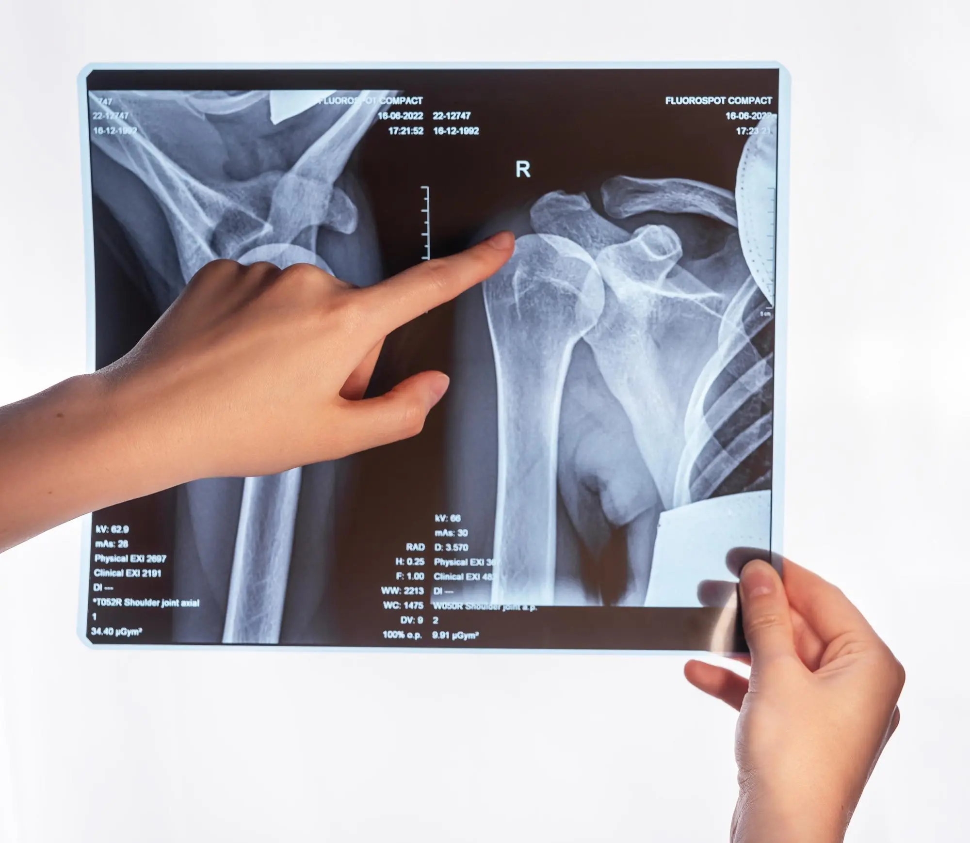
In the year 1895, Wilhelm Roentgen discovered Roentgen Rays, or X-rays as they’re more popularly known. The discovery was monumental for the world of medicine. So much so that an entire branch of medicine, radiology, was founded based on Roentgen’s discovery.
Radiology refers to the medical discipline that uses medical imaging modalities to visualize and thereby treat diseases and ailments within the human body. The technology is in no way limited; in fact, you can visualize the internal systems and working of nearly everything based on the penetrative and absorptive capacities of the test.
However, Roentgen only paved the way for other discoveries to be made in the world of radiology. It was soon discovered that the powerful rays generated by X-ray machines were disruptive to the body’s internal workings. To simplify, molecules within the body could not bear the strength of X-rays and would disintegrate.
It was quickly discovered that X-rays were doing more harm than good. This led researchers to discover other avenues to visualize the body’s inner workings; Magnetic Resonance Imaging.
What is an MRI?
Magnetic resonance imaging (MRI) is a medical imaging technique used to form images of the body’s anatomy and even visualize the biochemical and physiological processes that occur within it. MRI scanning does not involve the use of X-rays. Rather it incorporates the use of magnetic fields and gradients to generate images.
The ionizing potential with MRIs is significantly less compared to X-rays which makes it an ideal choice for medical imaging. These scans are typically used for:
- Neuroimaging
- Musculoskeletal imaging
- Angiography
- Cardiovascular imaging, and
- Imaging the gastrointestinal tract
The Procedure:
Compared to X-rays and computerized tomography (CT) tests, MRI scans do not utilize radiation. Radio waves simply re-adjust hydrogen particles that normally exist inside of the body. This doesn't create any molecular (ionized) changes in tissues.
As the hydrogen molecules get back to their standard arrangement, they transmit various levels of energy (based upon the sort of body tissue they are in). The scanner catches this energy and makes an image utilizing this data.
In most MRI units, the magnetic field is delivered by passing an electric flow through wire loops. Different loops are situated in the machine and are set around the part of the body being imaged. These loops send and get radio waves, delivering signals that are identified by the machine. The electric flow doesn't interact with the patient.
A PC measures the signs and makes a progression of pictures, every one of which shows a meager cut of the body. These pictures can be concentrated from various points by the radiologist.
Shoulder MRI Scan
Magnetic Resonance Imaging (MRI) scans of the shoulder are fairly common when it comes to musculoskeletal imaging. The modality is used commonly to assess bones, tendons, muscles, blood vessels, and injuries to any of them.
Normal shoulder MRI scans show properly aligned and non-fragmented bones, adequately working blood vessels, and no tears in muscles or ligaments.
Detailed Shoulder MRI with contrast allows doctors to examine the body and detect any disease. The images can be examined and assessed on a computer monitor. They may also be sent electronically, printed, or uploaded to a digital cloud server.
Related Test: Shoulder MRI without Contrast
Normal Vs. Abnormal Shoulder MRI
Your physician might advise an MRI for shoulder pain if the shoulder pain has manifestations indicative of any of the following conditions:
- Degenerative joint diseases (labral tears and arthritis)
- Fractures (commonly sustained in sports injuries)
- Rotator cuff disorders (impingements and tears)
- Joint abnormalities
- Osteomyelitis and other infections
- Benign or malignant tumours
- Bleeding within or inside of the joint
- Prognosis and progress after surgery
Shoulder MRI Cost:
Prices for MRI scans of your shoulder vary based on the location of the facility and the overall service it provides. Larger cities such as Los Angeles, Chicago, and Texas have more affordable rates because of competitive marketing.
Research hospital rates are fairly expensive and range between $3,000 to $6,000 per MRI, this might vary for normal shoulder MRI with contrast. The prices might seem hefty, but your insurance takes care of most of it:
- shoulder MRIs cost for a high-deductible insured patient at a large hospital: $3,000 - $6,000
- MRI shoulder cost for high-deductible insurance at imaging centre: $1,200 - $2,000
- MRI shoulder cost for a fully insured patient at a hospital (15% copay): $450 - $900
- MRI shoulder cost for a cash-paying patient at an imaging centre: $250 - $500
Risks of a Shoulder MRI:
The MRI exam poses minimal to no risk provided that appropriate safety guidelines and measures are followed. Some of which include:
- Use the appropriate amount of anesthesia. Too much of it might be harmful to the patient.
- Implanted medical devices might hinder the magnetic images.
- Nephrogenic systemic fibrosis is a recognized, but rare, complication related to the injection of gadolinium contrast.
- Allergic reactions are rare.
Conclusion
Magnetic Resonance Imaging (MRI) scans are common radiological modalities advised by physicians to visualize the body’s internal systems and work. Shoulder MRI scans (including MRI with arthrogram for shoulder pain, normal shoulder MRI with contrast, and MRI arthrogram shoulder labral tear) are commonly advised if your doctor suspects a fracture, an infection, trauma, or a tumor in your shoulder.


