
According to a Statista report on Medical Technologies, the United States has the highest MRI units per million population anywhere in the world. Around 40.44 MRI units are available per unit million in the United States.
Magnetic Resonance Imaging (MRI) is a radiological modality that was first introduced in 1977. The modality revolutionized the world of internal bodily imaging by being a safer option than X-rays (which radiology was heavily dependent upon up until the late 1900s).
MRIs do not necessitate the use of ionizing radiation as X-rays. Instead, a magnetic field is employed that ‘charges’ protons in the human body and via the transmitted field creates an image onto a computer screen.
MRIs are particularly helpful in understanding soft tissue functioning. Thereby, making MRI’s the first imaging modality advised by physicians for studying and examining normal and abnormal brain functioning.
The First Cranial MRI
The idea that every atom behaves as an individual magnetic field had been theorized and studied long before 1977 when the first MRI scan was performed on a live patient. The idea, however, was never employed outside of theoretical concepts and certainly not for medical technologies.
Isidor Issac Rabi theorized that the orientation of magnetic moments of nuclei could be aligned by electromagnetic waves of ample frequencies. Rabi’s theory was based on the existing belief that magnetic moments could be aligned either parallel or anti-parallel to external magnetic fields.
In 1937, Rabi extensively studied his theory and further inoculated into it that the same amount of energy that is given to an atom could be emitted back by the atom to a lower energy orientation. This was referred to as the molecular beam magnetic resonance.
MR Images of the Brain:
- The first MR images of the human brain were obtained in 1978 by researchers at EMI Laboratories.
- MRI angiography was first introduced by General Electric in 1986.
- The idea of contrast imaging in perfusion MRI was introduced in 1988.
- Shortly before Rabi died in 1988, he was imaged in an MRI machine. ‘It was eerie,’ he said, ‘I never thought my work would come to this.’
- Functional Magnetic Resonance Imaging (fMRI) was first introduced by AT&T Bell Labs by Seji Ogawa who recognized that oxygen-depleted blood was attracted in a magnetic field with dHb.
What is Magnetic Resonance Imaging of the Brain
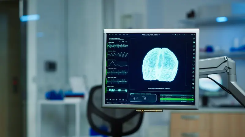
Magnetic Resonance Imaging (MRI) employs the use of a large donut-shaped magnet that encircles the patient. The magnet could also be described as a tunnel. Patients are asked to lie down on a table that slides into the tunnel.
Scans of the brain are painless, safe, and non-invasive tests for studying (and thereby diagnosing) pathological conditions of the brain. Alternatively, they could be used to rule out any pathology.
Physicians would often advise MRI scans over CAT scans and other X-ray scans because MRI scanners use radio waves instead of ionizing radiation rays. This means that MRI scanning is less hazardous (negligible compared to X-rays), but also more technique sensitive, and costly.
Radio waves basically manipulate the magnetic positions of the atoms inside of the body. The disruption is recorded and projected onto a computer screen. The computer is booted with an algorithm that calculates and then deduces the imaging in clear, cross-sectional, images of the brain.
Brain Conditions Diagnosed With MRIs:
An MRI scan can help in understanding the normal physiology and pathology of the human brain. Some pathological conditions commonly studied on MR images of the brain include:
- Infections
- Tumors
- Cysts
- Aneurysms
- Spinal cord injuries
- Stroke
- Multiple sclerosis
- Hydrocephalus
- Swellings
- Hormonal disturbances
- Inflammation
- Bleeding
- Prognosis of the previous injury
- Developmental issues
Preparing For a Brain MRI
- The technician would have you fill out a form to indicate any metal prosthetics that you are currently using and advise you to remove any objects that contain metal.
- If you have metallic prosthetics that cannot be removed in any case, you might not be eligible to have an MRI scan done.
- The technician or physician would go over the procedure with you and indicate that for obtaining the highest quality results, you would have to lay perfectly still.
- Some patients experience claustrophobia when they’re asked to lay still in an MRI scanner and might cause panic. These patients are often sedated or advised against getting an MRI scan.
- If the patient has to be sedated before the MRI scan, they will be advised to fast a few hours before the actual scan. This is to ensure that the IV sedative does not cause any untoward effects.
MRI Head Procedure
The procedure takes anywhere between half an hour to three-fourths of an hour where the patient is asked to stay as still as possible. A coil (plastic headgear) might be placed around the patient’s head. As the magnet rotates around the table, the technician will take images of the head that will be projected onto a computer screen.
Several different images in varying spatial rotations are taken. Each lasting for a few minutes.
How Much Does a Brain MRI Cost?
Brain MRI scans usually cost between $250 up to $2500 in the United States. The cost of an MRI scan is variable between one location and state, and the other. Freestanding clinics and imaging centers are often more cost-effective than hospitals and standalone clinics. Freestanding clinics are also better suited for insured patients with high deductibles.
When Would Your Physician Recommend an MRI
Your physician would recommend an MRI if he or she suspects any underlying pathology. Advising an MRI scan often comes after routine investigations and clinical procedures indicate a pathology.
Our clinical experts continually monitor the health and medical content posted on CURA4U, and we update our blogs and articles when new information becomes available. Last reviewed by Dr.Saad Zia on April 2nd, 2023.