For Physicians
Sign Up 0    

Myocardial Infarction (Heart Attack)

Overview

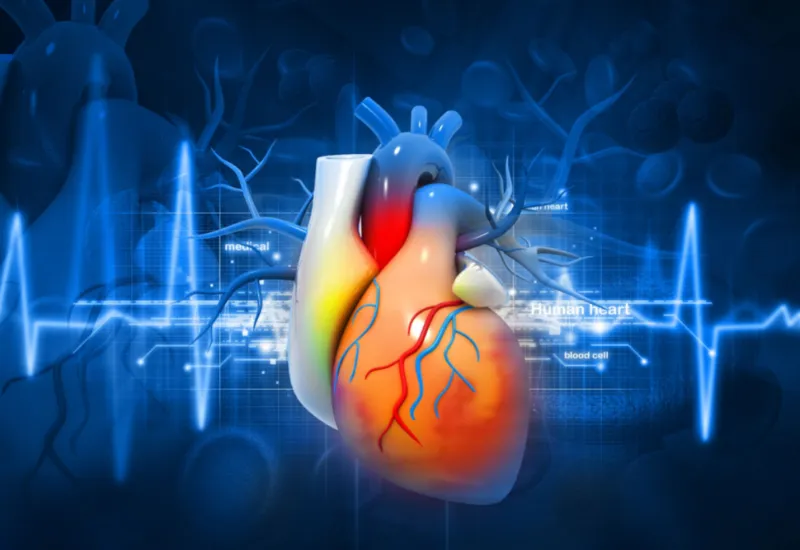
Myocardial infarction (MI), commonly known as heart attack, results when there is necrosis or irreversible death of heart muscle due to lack of oxygen to that muscle. Lack of oxygen occurs when there is an imbalance in the demand and supply of oxygen. The oxygen supply is interrupted as the coronary arteries are narrowed due to plaque rupture and thrombus formation.

The severity of MI or heart attack depends upon the duration and extent of ischemia (lack of oxygen) and the type of heart muscle involved. It is one of the leading causes of death in the world. In the United States alone, there are 1.5 million cases of myocardial infarction reported per year.

Causes

The majority of cases of MI are caused by atherosclerosis. Atherosclerosis is defined as the thickening of the vessel wall due to the build-up of cholesterol inside the lumen. The build-up can form plaque that ruptures and obstructs the lumen of the entire vessel. Atherosclerosis can be caused by various reasons such as high cholesterol levels in the body, high blood pressure, etc.

Nonatherosclerotic causes are the conditions or diseases that involve coronary vessels or cardiac muscles. These include certain drugs, myocarditis, coronary trauma, etc.

Types

MI can be divided into several types depending upon the underlying cause:

  • Type 1- spontaneous MI in which the symptoms develop spontaneously without any underlying obstructive coronary artery disease
  • Type 2- MI results from ischemia due to an imbalance between oxygen demand and supply. The imbalance includes coronary artery spasm, coronary artery embolus, endothelial dysfunction, arrhythmias, anemia, respiratory failure, hypotension, and hypertension.
  • Type 3- MI results in sudden death in the non-availability of cardiac biomarkers. It could be due to the reason that cardiac biomarkers could not be drawn from blood, or they were not released yet in the blood.
  • Type 4a- MI is related to the percutaneous intervention (PCI) in which the blockage in coronary arteries is evident from imaging along with loss of viable myocardium and wall motion defects.
  • Type 4b- MI related to stent thrombosis
  • Type 5- MI related to coronary artery bypass graft (CABG)

Another classification is based on the types of acute coronary syndromes in which there is a sudden loss of oxygen supply to the cardiac muscle resulting in ischemia or necrosis. These types include:

  • ST-elevation MI (STEMI)- ECG waves, in this case, show ST-segment elevation
  • Non-ST elevation MI (NSTEMI)- ECG waves do not show ST-segment elevation

Risk Factors And Epidemiology

Myocardial infarction is prevalent worldwide. It is one of the leading causes of death worldwide. In the United States, one-third of all deaths in the population older than 35 are due to MI.

Mortality associated with MI has decreased significantly due to better lifestyle choices, health-seeking behaviors, and education about the disease.

Risk factors for MI include:

  • Male gender
  • Middle age
  • Smoking
  • Hypercholesteremia
  • Family history of cardiac diseases
  • Hypertension
  • Diabetes
  • Obesity
  • Stress
  • Sedentary lifestyle
  • Coronary trauma
  • Drugs such as cocaine and amphetamine
  • Heavy exertion
  • Severe anemia
  • Respiratory infections

Signs And Symptoms

Following are the signs and symptoms of myocardial infarction:

  • Characteristic chest pain lasting for about 30-60 minutes, sharp, continuous, and intense in character. It radiates to the lower jaw, left arm, shoulder, and back. In some patients, it can be in the epigastric region mimicking acidity.
  • Sweating
  • Shortness of breath
  • Nausea or vomiting
  • Fatigue
  • Malaise
  • Increased heart rate
  • Increased rate of breathing
  • Increased blood pressure. In right ventricular MI, blood pressure is decreased
  • Coughing, wheezing may also occur

Complications of MI include:

  • Arrhythmias
  • Ventricular septal defects
  • Left ventricular aneurysm formation
  • Pericarditis
  • Pseudoaneurysm
  • Left ventricular mural thrombus

Diagnosis

 An ECG makes the diagnosis of myocardial infarction. In more than 80 percent of cases, it is confirmatory. Diagnosis is aided by specific laboratory tests that also help manage MI. These include:

  • Cardiac enzymes
  • Troponin levels are extremely sensitive for the cases of acute MI if performed within a few hours.
  •  Complete blood count
  • Liver function tests
  • Renal function tests
  • Serum electrolytes
  • Lipid profile
  • Imaging studies such as echocardiography and angiography can be used later to rule out coronary artery disease

Differential Diagnosis

Differential diagnoses of MI include:

  • Acute gastritis
  • APD
  • Aortic dissection
  • Aortic stenosis
  • Depression
  • Cardiogenic shock
  • Pneumonia
  • Gallstones
  • Infective endocarditis
  • COPD
  • Asthma
  • Pulmonary embolism

Treatment

  • Initial management of MI focuses on:
  • Supplemental oxygen supply
  • Immediate administration of aspirin
  • Pain relief with nitroglycerin
  • ECG

Once the patient is hospitalized, the initial management is continued with additional therapy that includes:

  • Continuous oxygen supplementation with oxygen saturation monitoring to maintain oxygen levels at greater than 90%
  • Additional pain relief with stronger analgesics such as morphine
  • Serial ECGs to track changes
  • Reperfusion through early intervention by the percutaneous intervention (PCI). It is associated with fewer chances of long-term complications and death.
  • Fibrinolysis with fibrinolytic agents such as streptokinase to achieve reperfusion.
  • Anticoagulation with heparin
  • Antiplatelet therapy with aspirin or clopidogrel
  • In cases where PCI fails, coronary artery bypass graft (CABG) may be indicated for long-term management.

Prognosis

The prognosis of myocardial infarction depends upon the time elapsed between the episode and treatment. It is associated with 30% mortality, and almost 50% of deaths occur before bringing them to the hospital.

If reperfusion treatment is started within 6 hours of the onset of MI, the prognosis is usually good, depending upon other factors such as comorbidities, age, and weight. When performed within 6 hours of the onset of MI, revascularization procedures can be most beneficial, preventing remodeling of the heart muscle and other associated complications.

Lifestyle Modifications

  • The most important prevention strategy against MI is adopting a healthy lifestyle so avoidable causes such as obesity and hypercholesteremia can be controlled.
  • People with a family history of cardiac diseases and hypercholesteremia should undergo screening for risk factors early and adopt lifestyle changes instantly to avoid MI.
  • Comorbidities such as hypertension and diabetes increase the risk of developing an MI. They should be kept in check and under control with proper treatment and healthy lifestyle choices.
  • Stop smoking.
  • Limit alcohol intake.
  • Recreational drugs such as cocaine and amphetamines should also be avoided.
  • Reduce daily stress by seeking help for stress management.

Our clinical experts continually monitor the health and medical content posted on CURA4U, and we update our blogs and articles when new information becomes available. Last reviewed by Dr.Saad Zia on May 25, 2023.


References

Heart failure after myocardial infarction: incidence and predictors - Jenča - 2021 - ESC Heart Failure - Wiley Online Library

https://onlinelibrary.wiley.com/doi/full/10.1002/ehf2.13144

Myocardial Infarction - StatPearls - NCBI Bookshelf (nih.gov)

https://www.ncbi.nlm.nih.gov/books/NBK537076/#:~:text=Myocardial%20infarction%20(MI)%2C%20colloquially,hemodynamic%20deterioration%20and%20sudden%20death.

Related Blogs

-->