Supraventricular tachycardia (SVT)

Overview

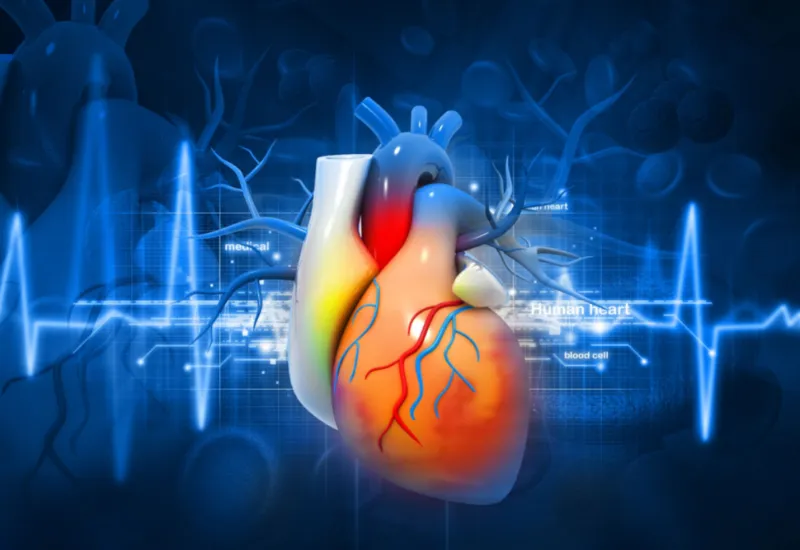
Supraventricular tachycardia (SVT) refers to the conditions associated with episodes of fast heartbeat occurring due to the problems of electrical activity in the upper chambers of the heart. A normal heart usually beats at the rate of 60 to 100 beats per minute. When the heart rate is more than 100, it is known as tachycardia. An episode of SVT can make the heart beat at the rate of 150-220 beats per minute. Your heartbeat may be so fast that it won't allow the heart to be filled with blood, resulting in insufficient blood supply to the body. You may feel palpitation, lightheadedness, faintness, shortness of breath, sweating, and/or chest pain.

Causes

Your heart consists of four chambers, the upper two are called atria, and the lower two are called ventricles, which contract at a regular rhythm to supply blood to the body. This rhythm is generated and controlled by a special electrical system in your heart consisting of the SA node, AV node, and bundle of hiss. Any problem occurring in the conductivity of this electrical system can cause abnormal rhythms known as arrhythmias. SVTs that arise and contract the atria are usually due to re-entry of the electrical impulses through another pathway or increased automaticity. These impulses can also be transferred to the lower chambers, causing premature beats in the ventricles.

SVT may occur in a healthy person or people with concurring diseases. The factors that can trigger an SVT may include;

·         Previous or current heart disease such as heart attack, rheumatic heart disease, mitral valve prolapse, pericarditis, Heart failure, Wolff-Parkinson-White syndrome

·         Chronic lung disease or pneumonia,

·         Excessive intake of caffeine, alcohol

·         Drug abuse, including methamphetamine and cocaine

·         Pregnancy

·         Smoking

·         Thyroid disease

·         Digoxin toxicity

·         Some medications, such as asthma medications, cold and allergy drugs

Types

Supraventricular tachycardias usually arise from the SA node, atria, or AV node and are classified by their specific site of origin.

Sinoatrial origin:

1.       Sinoatrial nodal reentrant tachycardia (SNRT)

Atrial origin:

1.       Ectopic atrial tachycardia (EAT)

2.       Multifocal atrial tachycardia (MAT)

3.       Atrial fibrillation with rapid ventricular response

4.       Atrial flutter with rapid ventricular response

Atrioventricular origin:

1.       AV nodal reentrant tachycardia (AVNRT)

2.       AV reciprocating tachycardia (AVRT) – (including Wolff-Parkinson-White syndrome)

Risk Factors And Epidemiology

Some of the conditions that can predispose you to get an SVT include;

·         Age: Children and females

·         Anxiety disorder

·         Excessive alcohol, tobacco, or caffeine use

·         Strenuous exercise

·         Long-standing Diabetes mellitus or hypertension

·         Heart or lung disease

·         Hyperthyroidism

·         Obstructive sleep apnea

In the USA, paroxysmal SVT affects around 0.2% of the population with a frequency of approximately 1-3 cases per 1000 people. The prevalence of Atrial fibrillation is 0.4-1% in the population, making it the most common arrhythmia. Females are affected more. The chances of SVT Increase with age.

Signs And Symptoms

The presence of the following symptoms may point toward an SVT, and you should plan to consult a physician.

  • Palpitation
  • Feeling faint, vertigo, and collapse
  • Chest pain
  • Fatigue
  • Shortness of breath
  • Breathing difficulty on lying flat
  • Irregular heartbeats

Diagnosis

To diagnose the disorder, your doctor will inquire about the symptoms and the previous medical records. They will examine you with particular emphasis on the heart examination using a stethoscope for hearing the heart sounds and murmurs. The following tests can be used;

·         Electrocardiogram: Electrical impulses in the heart are recorded during this simple test. Many cardiac disorders, such as irregular heartbeats and clogged arteries, can be diagnosed with an ECG. ECG is sometimes taken when you work out, usually on a bike or treadmill. A stress test is a name for this approach.

·         Ambulatory monitoring: It makes use of sensors that monitor your heart rate. A Holter monitor is a device that is worn for one or more days to record the activity of your heart.

·         Event recorder: This device can be worn even for a month to detect infrequent arrhythmias. You are required to press a button whenever you feel the symptoms.

·         Electrophysiology: During this procedure, a long, narrow, flexible tube is placed into a blood channel generally in the pelvis and directed to the heart to detect the electrical activity of the heart through sensors located at the tip of the catheter. It can help a doctor locate the accessory pathways.

·         Echocardiogram: In this test, ultrasound waves create an image of your heart. This test would be suggested by your physician to check how your heart's structures work.

·         Blood tests: Various blood tests, such as those to assess your kidneys, liver, thyroid function, and digoxin levels, may be ordered.  

·         Cardiac enzymes: Increased amounts of specific proteins or enzymes found in cardiac muscle may be detected by blood testing. Any person with chest pain and risk factors should be checked for myocardial infection using a blood test for enzymes.


·         X-ray of the chest. A chest X-ray can reveal the state of the lungs, as well as the size and form of the heart and main blood channels. A chest X-ray can also show lung disorders such as pneumonia or a collapsed lung that can predispose to SVT.

Differential Diagnosis

Some other disorders may present like SVT, that need to be excluded to make an efficient diagnosis;

·         Thyrotoxicosis

·         Pregnancy

·         Cocaine Toxicity

·         Congenital heart diseases

·         Myocarditis

·         Valvular heart disease

·         Heart Failure

·         Infective Endocarditis

Treatment

Some people with no symptoms usually do not need urgent treatment. The goal of the treatment is to control the rate and prevent hemodynamic collapse. The following treatment is usually performed;

Vagal Maneuvers: When the patient is stable, vagal maneuvers can be tried. They stimulate the vagal nerve and release a chemical that helps to slow down the heart rate. Examples of such maneuvers are breath-holding, carotid massage, and the Valsalva maneuver (it increases air pressure in your chest. You need to breathe out strongly through your mouth, keeping your nose tightly closed).

Electrical cardioversion: It is the first choice for unstable patients. During this procedure, an electric current is delivered to your chest using special paddles to restore electrical activity.

Medications: Medications are used to boost blood flow and control your blood pressure. Your doctor will prescribe you medication to decrease your heart rate, like Beta Blockers (Atenolol, Propranolol) and Calcium Channel Blockers (Amlodipine, Diltiazem ).

Pacemaker: Some people are given a pacemaker device implanted under the skin. It is connected to the heart and generates a heartbeat.  


An implantable cardioverter-defibrillator (ICD): This equipment measures your heart rate and, if necessary, gives electric shocks to correct irregular heart rhythms. It monitors and manages aberrant arrhythmias.

Prognosis

The outcome depends on the frequency and severity of SVT. Some patients may not have symptoms, while some can show hemodynamic instability. Some SVTs can lead to ventricular fibrillation and sudden death.  

Prevention

Adopting the following lifestyle changes may prevent you from developing valvular heart disease.

  • Quit smoking
  • Take a healthy balanced diet and avoid oily fried food.
  • Perform some kind of exercise regularly.
  • Keep your health status in check by regularly getting assessed for Diabetes Mellitus, high blood pressure, etc.
  • If you are suffering from any risk factors, keep it controlled very well by adhering to its treatment. 

Our clinical experts continually monitor the health and medical content posted on CURA4U, and we update our blogs and articles when new information becomes available. Last reviewed by Dr.Saad Zia on April 29th, 2023.

References

https://www.mja.com.au/system/files/issues/190_05_020309/med10727_fm.pdf

https://www.nhs.uk/conditions/supraventricular-tachycardia-svt/

Related Blogs